FLIR Thermal Science Cameras Support Medical Research
- 11 Dec 2020
Renowned US University uses FLIR cameras to study the effect of temperature on tissue autofluorescence.
Tissue autofluorescence spectroscopy is a technique that measures the intrinsic light emissions from biological agents upon excitation by a light source. It has been demonstrated in a variety of biomedical research for differentiating between normal and diseased tissue, however, the relationship between autofluorescence and temperature has not been well defined. Currently, researchers at a renowned US university are conducting experiments with the help of a thermal imaging camera from FLIR systems to better understand this relationship.
Autofluorescence spectroscopy can be used for the purpose of surgical guidance, for example as an imaging technique for delineating surgical margins during cancer excisions (e.g. to be able to identify cancerous tissue close to the margin of normal tissue). As such, by studying autofluorescence, universities are trying to bridge the gap between fundamental science and their practical applications to healthcare.
Effect of Temperature on Autofluorescence
Despite its use in biomedical research, the relationship between autofluorescence and temperature in tissue has not been explicitly defined. Many past studies have often assumed a constant temperature during measurements where in vivo experiments were assumed to be at body temperature and in vitro experiments were assumed to be at room temperature. However, there are instances in which the temperature may vary greatly such as during ablation surgeries. In these cases, an understanding of temperature effects on tissue autofluorescence is potentially crucial for accurate interpretation of results during these procedures.
In a series of preliminary experiments aided by the use of a FLIR A655sc thermal imaging camera, researchers from a renowned US university have shown an inverse relationship between temperature and autofluorescence for ex vivo human tissue.
Since the researchers were correlating the effects of temperature to our recorded autofluorescence signals, it was very important to acquire accurate and reliable temperature values.
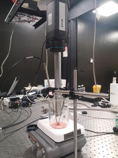
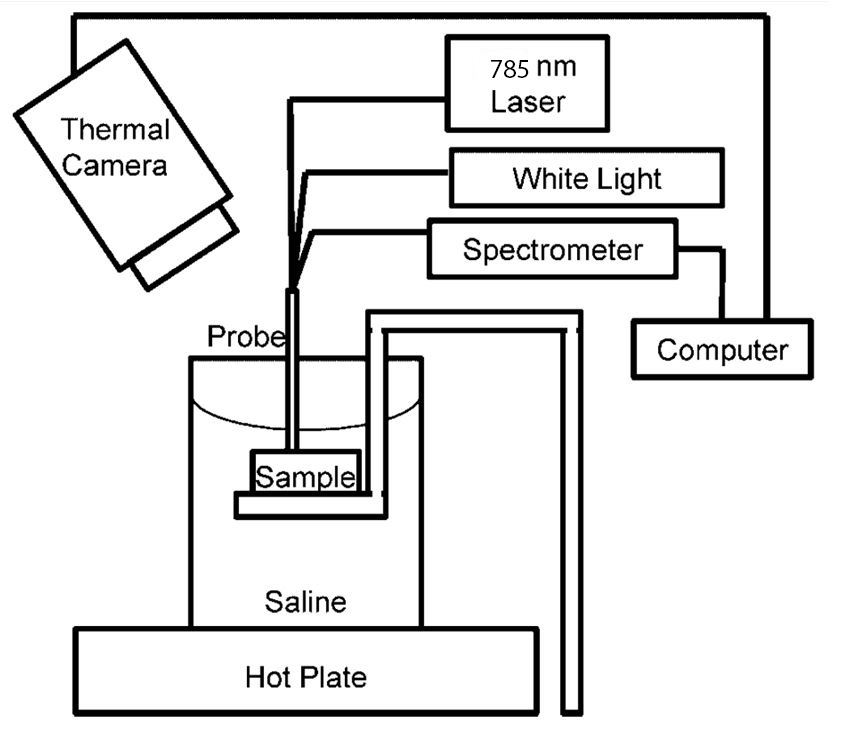
Test set-up
A spectroscopy system equipped with a dual-excitation probe was used to measure the autofluorescence and diffuse reflectance of ex vivo human muscle tissue. Excitation light was provided by a 785 nm diode source while near-infrared autofluorescence emissions were collected by a charge coupled device (CCD) at a spectral resolution of 3.15 nm. The tissue’s temperature was continuously measured by a FLIR A655sc thermal imaging camera.
Ex vivo human muscle samples were acquired and stored at -80°C. During experiments, the samples were placed into a glass beaker and measurements were taken as the samples were allowed to passively warm up to room temperature. Once the sample reached 20°C, the sample was then submerged saline and gradually heated by a hotplate with measurements recorded at every 5°C increase in temperature.
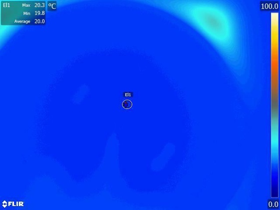
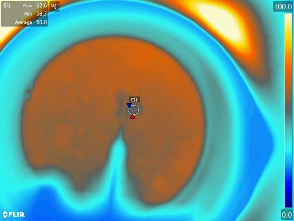
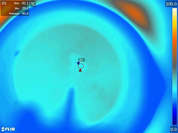

Figure: (a-c): Wide-field temperature maps recorded from ResearchIR at 25°C, 40°C, and 60°C. Circle region of interest is placed on top of tissue sample. (d.) Plot of 833 nm fluorescence emission over time. Note the decrease in fluorescence intensity as temperature increases.
Reliable temperature values
"Since we are correlating the effects of temperature to our recorded autofluorescence signals, it was very important for us to acquire accurate and reliable temperature values," a project researcher comments. "We chose to use the FLIR A655sc thermal imaging camera because of its affordability as a qualitative temperature imaging tool. In addition, we were able to equip the camera with a variety of magnification lenses to better view our small tissue samples."
The FLIR A655sc thermal imaging camera is a high-resolution (640 x 480 LWIR with 17 micron pixels) thermal imaging camera with an uncooled detector that is specifically aimed at scientists and researchers. The camera has Gigabit Ethernet and USB outputs, a wide variety of lens options, and different mounting options, which makes it easy to use in combination with microscopes.
The researchers involved in the project say that these experiments were their first time using a FLIR thermal imaging camera. They said they were surprised how easy this camera was to use. "You just plug it in, connect it with the software, and you're ready to go."
In an experiment with ex-vivo human tissue, a dualexcitation probe-based system was used to measure fluorescence and diffuse reflectance.
IR Camera Control and Analysis Software
During the test set-up, the researchers also used FLIR’s ResearchIR software to view the live IR image, change camera settings, record single images, make video recordings, and analyze the temperature data in the images. The ResearchIR software connects directly to FLIR Research and Science cameras via USB, Firewire, Gigabit Ethernet, and Camera Link to acquire thermal snapshots or movie files. The software can perform real-time image analysis, with an extensive set of measurement tools including spots, lines, and areas.
A researcher from the test team commented: "I found the FLIR ResearchIR software very easy to set up and intuitive to use. I really liked how I could define different regions of interest and shapes for real time temperature feedback. This was particularly helpful during the experiment."
Notice of Intended Use: FLIR Systems, Inc. does not intend for the FLIR A655sc thermal imaging camera to be used as a medical device. The camera is not intended to be used for any diagnostic, therapeutic, or preventative purposes for humans or animals, nor is intended to be used to affect the structure or function of the body of a human or an animal. Any such use of the FLIR A655sc is not authorized by FLIR Systems, Inc.


